PGD strategy characterised by a nested-PCR protocol producing amplicons analysable by the minisequencing technique (Fiorentino et al., 2003) is used or B-Thalassemia (Figure 1), thus avoiding the use of mutation-based strategies that require optimisation of specific PCR protocols for each mutation to be analysed. The minisequencing approach has proven to be extremely flexible and appropriate for the analysis of a wide spectrum of mutations and compound genotypes. Although each PGD case can involve the presence of different mutations and genotype combinations, the use of a panel of PCR primers tested preclinically enables a substantial shortening of the preliminary phase for each couple.
Figure 1.
Preimplantation genetic diagnosis (PGD) for B-thalassemia performed by using the minisequencing technique.
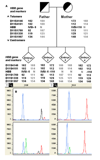
A) Pedigree of a couple carrying B-thalassemia mutations and examples of different results of the HBB gene mutation analysis. Informative STR markers are ordered from telomere (top) to centromere (bottom). The numbers in STR markers represent the size of PCR products in base pair. STR alleles linked to the paternal and maternal mutations are represented in boldface.
B) Examples of minisequencing results obtained for the above case. The mutations of interest are IVSI-110 G-A and IVSI-6 T/C, analysed in a multiplex reaction format. The y-axis represents the Relative Fluorescence Units (RFU) of the detected fragments; the x-axis represents time and is displayed by data points. Colour is assigned to individual ddNTPs as follows: green/A, black/C, blue/G, red/T. Mutation IVSI-110 G-A is shown on the left of the minisequencing window; the blue peak represents the normal allele (wild type base G), the green peak (mutant base A) the mutated allele. Mutation IVSI-6 T/C is shown on the right; the red peak is the normal allele (wild type base T) and the black peak (mutant base C) is the mutated allele.
Embryo 1 (upper panel/left) is carrier for IVSI-6 T/C mutation; Embryo 3 (upper panel/right) is normal; Embryo 2 (lower panel/left) is compound heterozygote for the two mutations. Embryo 4 (lower panel/right) is also affected, although minisequencing result shows a heterozygosity for mutation IVSI-110 G-A. In fact, linked STR markers highlight an allele drop-out (ADO) of the affected allele (black peak, mutant base C).