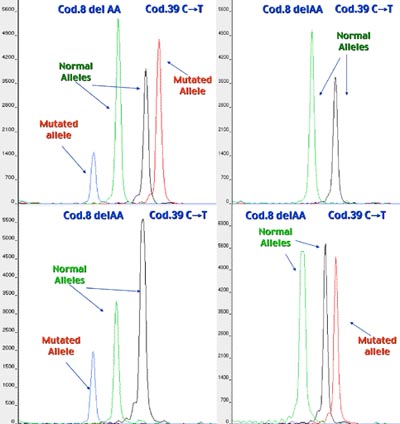
Currently, PGD for single gene disorders can be accomplished by either polar body removal or blastomere biopsy. Which method may be used is determined on a case-by-case basis. This procedure can be used to select for embryos that do not have a specific genetic disease by testing the polar bodies or blastomeres for the genetic mutation.

Inside the test tube containing the blastomere, a solution is added that permits cell lysis and thus the liberation of DNA from the cell nucleus. Subsequently, by means of in vitro enymatic ampification known as the Polymerase Chain Reaction (PCR), the genetic region of interest, involved in the mutations that is being searched for, is amplified millions of times. The genic amplification product then undergoes mutation analysis to search for genic mutions present in the couple. The analysis of mutations is the most important and delicate phase of PGD.
To guarantee maximum interpretative reliability, it is indispensable that it be carried out with methods and instruments that permit the univocal identification of the mutations that are being searched for. Automated sequence analysis is presently the best method of genetic analysis, inasmuch as it permits the exact determination and direct visualization of a specific mutation. The application of this technique to PGD is done by the use of completely automated state of the art equipment.

PGD centers worldwide are concerned about the possibility of misdiagnosis, which can occur as a result of failure of allele specific amplification, or allele dropout (
ADO). In basic terms, this is the failure of one of the genes (allele) to show up in the analysis (it “drops out” of the picture.) ADO is of concern primarily in blastomere biopsy when each parent carries a different mutation for a recessive condition.
To minimize the potential for diagnostic error by PGD, it is preferable to offer polar body removal or to perform a combined analysis of the specific mutation and specific linked markers that are inherited along with the gene. This dual amplification allows for higher accuracy and for the detection of ADO, thus enhancing diagnostic accuracy and reducing the risk for misdiagnosis.